Plasmid Name
pHC-IgG1-LALA
Mutation Set
L234A + L235A (Lower Hinge)
Selection Marker
Ampicillin
Promoter
CMV
Fc Engineering Overview
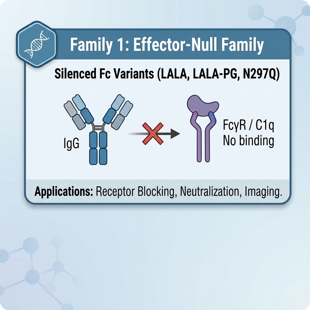
The human IgG1 (LALA) Fc variant introduces two alanine substitutions (L234A/L235A) into the lower hinge region to disrupt the canonical hydrophobic interaction interface required for FcγR engagement and complement C1q recruitment. It significantly reduces immune effector functions while preserving native endosomal FcRn recycling, structural stability (Tm ≈ 70°C), and normal IgG1 pharmacokinetics. LALA is the foundational benchmark legacy effector-null Fc variant widely used in therapeutic antibody discovery.
Functional Profile
| Property | Effect |
|---|---|
| FcγRI (CD64) binding | Residual Low-Affinity Binding Retained ↓↓↓ |
| FcγRIIa/IIb/IIIa binding | Severely Reduced / Minimal ↓↓↓ |
| ADCC | Strongly Attenuated / Minimal ↓↓↓ |
| ADCP (Macrophage Phagocytosis) | Strongly Attenuated / Minimal ↓↓↓ |
| C1q binding | Strongly Reduced ↓↓ |
| CDC | Strongly Reduced ↓↓ |
| FcRn binding & half-life | Normal / WT Baseline |
| Glycosylation & Thermal Stability | Intact (N297 glycan retained; native CH2 stability Tm ≈ 70°C) |
Mechanism of Action
- Lower Hinge Disruption: Residues L234 and L235 form a vital hydrophobic contact patch within the lower hinge (EU 231–236). Replacing these hydrophobic leucine residues with alanine severely destabilizes binding across the FcγR family.
- C1q Depletion: The mutations partially collapse the adjacent hinge-CH2 junction, reducing initial C1q attachment and subsequent complement cascade progression.
- Residual Binding Note: Because the proline-sandwich residue (P329) remains wild-type, LALA retains minor residual binding to high-affinity FcγRI (CD64), which can trigger subtle background activation under high receptor density.
Applications
- Standard legacy baseline control for evaluating effector-attenuated antibody candidates.
- Checkpoint inhibitors (PD-1, PD-L1, TIGIT) requiring reduced effector engagement.
- Blockade and neutralization therapies where full effector activity is undesirable.
- Comparative benchmark against reinforced double-silent variants (LALA-PG, EN).
Plasmid Map & Feature Annotation
Insert Structure: VH – CH1 – hinge – lower hinge(L234A/L235A) – CH2(WT) – CH3(WT)
- VH: Variable heavy domain
- CH1: Constant heavy 1
- Hinge: Native IgG1 core hinge
- Lower Hinge (L234A/L235A): Double alanine substitutions at EU positions 234 and 235
- CH2(WT): Wild-type CH2 domain (retains native N297 glycan)
- CH3(WT): Native IgG1 CH3 domain
Product Note: While IgG1 (LALA) remains a widely published and historically validated effector-null backbone, developers seeking total elimination of residual FcγRI binding or CDC should consider the reinforced human IgG1 (LALA-PG) or human IgG1 (EN) variants.
EffectorNull Family Comparison Matrix
| Variant | Mechanism | Silencing Strength | Notes |
|---|---|---|---|
| human IgG1 (WT) | Wild-Type baseline | Baseline | Human WT Control. Native FcγR and C1q binding; fully active baseline control. |
| human IgG1 (EN / Armour) | Lower hinge & CH2 chimeric substitution | ★★★★★ | Ultra-Silent Chimeric Null. Cross-subclass IgG2/IgG4 sequence replacement; abolishes FcγR & CDC engagement. |
| human IgG1 (LALA-PG) | Lower hinge & CH2 proline-sandwich null | ★★★★★ | Clinical Gold Standard. Complete, double-silent suppression of all human FcγRs and C1q/CDC. |
| human IgG1 (LALA) | Lower hinge double alanine substitution | ★★★☆☆ | This Product. Classic Benchmark Control. Widely used legacy variant; retains minor residual FcγRI (CD64) binding. |
| human IgG1 (ΔG236) | Lower hinge single residue deletion | ★★★☆☆ | Selective FcγR-Null. Dislocates FcγR binding site while preserving native N-glycan structure and C1q loops. |
| human IgG1 (N297Q) | CH2 domain N-glycan site elimination | ★★★★☆ | Human Aglycosylated Control. Broad effector silencing via glycan removal; lowers thermal stability (Tm). |
Storage & Handling
- Plasmid: Store at −20°C
- Purified Antibody: Store at 2–8°C for short-term; −80°C for long-term. Avoid repeated freeze–thaw cycles.
- Use sterile techniques for all handling.
References
- Xu D, Alegre ML, Varga SS, et al. In vitro characterization of five humanized OKT3 effector function variant antibodies. Cell Immunol. 2000;200(1):16-26.
- Hezareh M, Hessell AJ, Jensen RC, et al. Effector function activities of a panel of mutants of a broadly neutralizing antibody against human immunodeficiency virus type 1. J Virol. 2001;75(24):12161-12168.